

Research Theme
Exploring the Evolutionary Origin of the Mammalian Neocortex
Understanding neuronal differentiation through development and evolution.
The mammalian neocortex, which constitutes a large part of the human cerebral cortex, is composed of diverse neuronal cell types organized into a characteristic six-layered structure. Interestingly, birds and reptiles—our fellow amniotes—possess neuronal cell types with properties comparable to those found in mammals, yet their pallial architectures differ markedly: the avian pallium is predominantly organized into nuclear structures, whereas the reptilian pallium exhibits a simpler, approximately three-layered organization. How, then, did these distinct brain architectures emerge despite the presence of broadly shared neuronal cell types?
To address this question, I study brain development across amniotes, particularly mammals (e.g., mice), birds (e.g., chickens), and reptiles (e.g., geckos). By comparing how neurons are generated, migrate, differentiate, and assemble into neural circuits during embryonic development, I aim to identify the cellular and molecular mechanisms that gave rise to the six-layered mammalian neocortex during evolution. Ultimately, this comparative approach seeks to uncover fundamental principles underlying the evolutionary diversification and increasing complexity of the vertebrate brain.
A particular focus of my research is cell lineage—the developmental relationship among cells derived from a common neural stem or progenitor cell. Lineage and clonal analyses provide powerful approaches for understanding how neural progenitors generate cellular diversity and how these cells subsequently contribute to brain architecture. Using genetic tools that I developed during my research in France, together with modern imaging, transcriptomic, and spatial analysis approaches, I investigate cell lineage and developmental dynamics across multiple amniote species.
Research Background
Undergraduate Research (1999–2003)
Kagoshima University, Faculty of Education
Science Education Program (Zoology)
As an undergraduate student, I studied animal behavior, focusing on behavioral observations of jumping spiders for my graduation research. During this period, I also obtained teaching licenses for elementary, junior high, and high school education.
Graduate Research (–2009)
The United Graduate School of Agricultural Sciences, Kagoshima University
Laboratory of Prof. De-Xing Hou
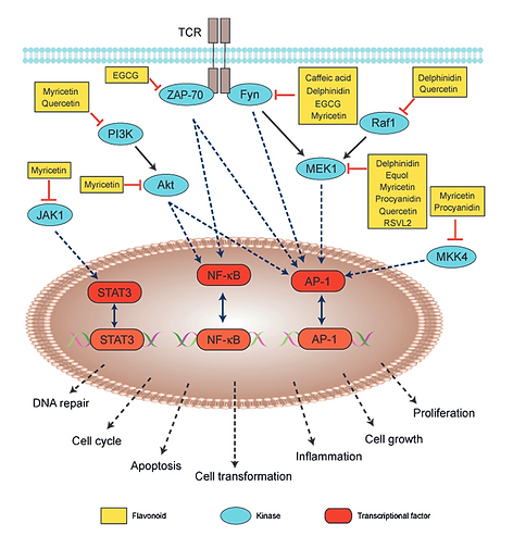
For approximately six years, from before entering the master’s program through completion of my PhD, I studied the molecular mechanisms by which dietary polyphenols, particularly flavonols, suppress cellular transformation.
Using cultured cell models susceptible to transformation, I investigated interactions between flavonols and intracellular signaling pathways. In particular, I developed an approach to identify protein kinases directly targeted by polyphenols after their uptake into cells, allowing us to investigate the primary molecular targets underlying their biological effects.
Postdoctoral Research I (2009–2015)
RIKEN Center for Developmental Biology (CDB), Kobe, Japan
Laboratory of Dr. Carina Hanashima
During my first postdoctoral position at RIKEN CDB, I moved into developmental biology and developmental neuroscience and began studying the mechanisms that control neural stem cell fate.
The diverse neuronal populations of the cerebral cortex are generated sequentially from common neural progenitor cells during embryonic development. This temporal regulation ensures that different neuronal populations are produced at appropriate developmental stages and in appropriate proportions. Understanding how neural progenitors switch from producing one neuronal subtype to another is therefore fundamental to understanding cortical development.
I focused on Foxg1, a transcription factor expressed in the developing telencephalon, and investigated its function during early neurogenesis. We discovered that Foxg1 acts as a key regulator of the developmental transition from the production of the earliest-born Cajal–Retzius cells to subsequently generated deep-layer cortical neurons.
Our genomic analyses further suggested that aspects of this Foxg1-dependent developmental program may have been modified during mammalian evolution. Because the six-layered neocortex is a defining feature of mammals, these findings led me to ask whether evolutionary changes in developmental programs could have contributed to the emergence of mammalian-specific cortical architecture.
This question became the foundation of my current research on the development and evolution of the vertebrate brain.
Postdoctoral Research II (2015–2020)
Institut de la Vision, Paris, France
Team Jean Livet
As I began extending my research from mouse cortical development to comparative studies of brain evolution, I encountered a major technical limitation. Genetic manipulation and lineage tracing are well established in mice, but comparable approaches are much more difficult in other vertebrate species, including birds and reptiles.
One important problem is episomal expression. Conventional plasmid-based expression systems can produce substantial transient expression from non-integrated DNA after transfection or electroporation, making it difficult to distinguish cells carrying stable genomic integrations from those showing only transient expression.
During my postdoctoral research in Paris, I therefore developed the integration-coupled On (iOn) switch, a genetic system designed to activate gene expression upon genomic integration. The iOn system makes it possible to identify stably integrated cells shortly after gene delivery and facilitates long-term labeling, lineage analysis, and genetic manipulation using somatic transgenesis.
This approach opened the possibility of applying genetic lineage and developmental analyses to species in which conventional transgenic approaches are difficult or impractical.
More information about the iOn system and related genetic tools is available on the Tools page.
Current Research (2020–Present)
Neural Circuit Development Project
Tokyo Metropolitan Institute of Medical Science, Tokyo, Japan
My current research integrates developmental biology, evolutionary neuroscience, comparative neuroanatomy, transcriptomics, spatial analyses, genetic manipulation, and imaging to understand how vertebrate brains acquire their species-specific architectures.
A major focus is the development and evolution of the cerebral cortex and pallium. By comparing mammals, birds, and reptiles, I investigate how conserved neuronal cell types are generated, migrate, differentiate, and become organized into markedly different brain architectures during development.
I am particularly interested in early-born neuronal populations, including subplate neurons and Cajal–Retzius cells, and how their developmental programs and functions have changed during vertebrate evolution. More broadly, I aim to understand how evolutionary modifications of developmental programs—including neural progenitor competence, neuronal migration, cell lineage, and neuron–glia differentiation—have contributed to the emergence and diversification of the mammalian neocortex.
Alongside these biological questions, I continue to develop and apply genetic tools for visualizing cell lineage, neuronal morphology, and developmental dynamics across species. These tools, including the iOn system, are also available for collaborative studies and distribution to the research community.
Published plasmids are available through Addgene. Please feel free to contact me if you are interested in these tools or in potential collaborations.


Neurons in the chick retina (RPE layer)
Three-color labeling using the LiOn switch



Three LiOn vectors (GFP, RFP, and iRFP) were introduced into the chick brain at embryonic day 4 (E4) and analyzed at E7.

